
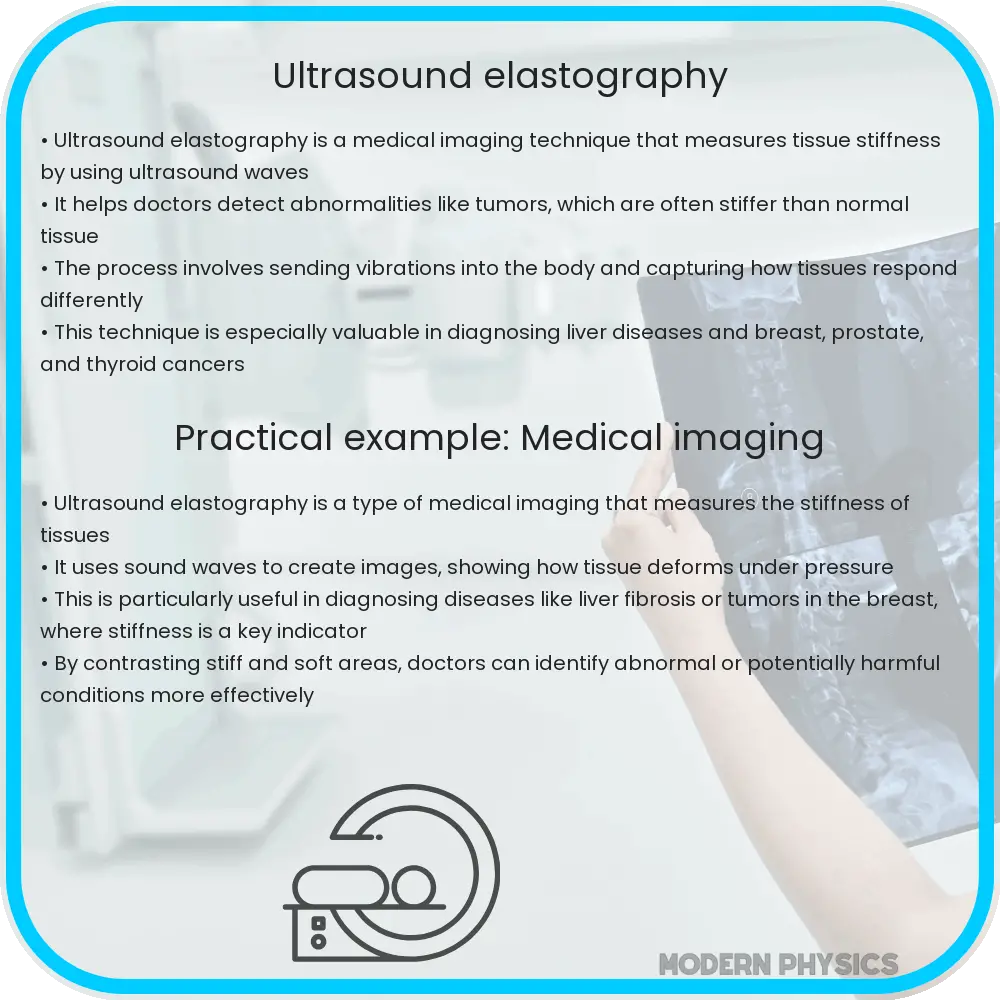
Ultrasound elastography is a non-invasive imaging technique that assesses the elasticity or stiffness of soft tissues, aiding in disease diagnosis and management.
Introduction to Ultrasound Elastography
Ultrasound elastography is a non-invasive imaging technique that maps the elasticity or stiffness properties of soft tissues in the body. This method is particularly useful in the medical field as it helps in the diagnosis and management of various diseases by providing additional mechanical property details that are not visible on conventional ultrasound. The technology leverages the principles of tissue displacement and mechanical stress to visualize how tissues deform under force.
How Ultrasound Elastography Works
At its core, ultrasound elastography works by applying a mechanical force, either externally or internally, to the tissue of interest. This force induces a displacement, which is then measured by ultrasound waves. Depending on the type of elastography used, this force can be generated through manual compression (static elastography) or acoustic radiation (dynamic elastography). The ultrasound machine captures the dynamic response of the tissue to this stress and compares it to scans without the applied force, allowing for the calculation of tissue stiffness.
The tissue’s response is measured in terms of strain, which is a relative change in dimension in response to stress. The general relationship between stress (σ) and strain (ε) in tissues can be expressed as:
σ = E * ε
Here, E represents the Young’s modulus, a measure of stiffness. Higher values of E indicate stiffer tissues. By analyzing these parameters, doctors can distinguish between healthy and pathological tissues, making it a powerful diagnostic tool.
Applications of Ultrasound Elastography
Ultrasound elastography has found applications across various areas of medicine. Some of its key uses are:
- Liver disease: It’s widely used to assess liver fibrosis, reducing the need for invasive liver biopsies.
- Breast cancer: It helps in differentiating benign from malignant breast lesions, thus aiding in cancer diagnosis and treatment planning.
- Thyroid nodules: Elastography can help in assessing the risk of thyroid cancers.
- Musculoskeletal conditions: It is used for assessing muscle and tendon integrity, particularly valuable in sports medicine.
Advancements in Ultrasound Elastography: Trends and Precision
As technology has advanced, so has the precision and utility of ultrasound elastography. Recent innovations have led to the development of techniques like shear-wave elastography (SWE), which provides quantitative measurements of tissue stiffness by measuring the velocity of shear waves through the tissue. This advancement allows for more standardized and reproducible assessments.
Furthermore, 3D elastography is merging the spatial mapping capabilities of three-dimensional ultrasound with elastographic techniques to provide detailed volumetric assessments of tissue stiffness. Such advancements have significantly increased the diagnostic accuracy and have paved the way for expanded applications in clinical practices.
Accuracy and precision in ultrasound elastography are continually improving with the development of sophisticated algorithms for image processing and enhancements in ultrasound technology. These improvements provide clearer, more detailed images and quantitative data, offering better insights into tissue health.
Precision in Practice
The precision of ultrasound elastography heavily relies on both the equipment used and the operator’s expertise. Factors such as probe pressure, patient movement, and the inherent heterogeneity of biological tissues can affect measurements. However, with proper calibration and standardized examination protocols, these challenges can be minimized, allowing for consistent and reliable use of ultrasound elastography in clinical settings.
Challenges and Future Directions
Despite its numerous benefits, ultrasound elastography faces certain challenges. Artifacts and noise can sometimes compromise the quality of the images obtained, potentially leading to diagnostic inaccuracies. Moreover, the interpretation of elastography results requires specific training and expertise, which can limit its widespread adoption in less-specialized settings.
Looking ahead, ongoing research aims to enhance the technology’s precision, ease of use, and application scope. Innovations such as artificial intelligence and machine learning are beginning to be integrated, offering the potential to automate aspects of the imaging and diagnostic process. This could lead to faster, more accurate interpretations and potentially broader global accessibility.
Educational and Training Aspects
As ultrasound elastography technology becomes more complex, the need for specialized training becomes evident. Educational programs and workshops designed specifically for this technology are crucial in equipping healthcare professionals with the necessary skills. Such training ensures that practitioners not only acquire theoretical knowledge but also develop practical skills in applying this technology effectively.
Universities and medical institutions are increasingly incorporating elastography into their curricula and continuous professional development programs. This initiative helps clinicians stay updated with the latest techniques and applications, ultimately enhancing the quality of patient care.
Conclusion
Ultrasound elastography has revolutionized the approach to diagnosing and managing various medical conditions by providing detailed insights into the mechanical properties of tissues. With applications spanning from liver disease to sports injuries, its importance in modern medicine continues to grow. The combination of ongoing technological advancements and enriched training programs promises to expand its capabilities and accessibility further. As such, ultrasound elastography stands as a pivotal example of how engineering and physics principles can be harnessed to improve clinical outcomes and patient care in the medical field.