
Explore the intricacies of compound microscope optics, including precision, magnification, and clarity, and how they unlock the microscopic world.
Understanding the Fundamentals of Compound Microscope Optics
The compound microscope, a cornerstone tool in scientific research, leverages the principles of optics to magnify small objects, enhancing the precision, magnification, and clarity of the images viewed. This intricate device, composed of multiple lenses, allows for the detailed examination of microorganisms, cells, and crystals that are invisible to the naked eye. Understanding how these components work together provides insight into the microscope’s capabilities and its application in various scientific fields.
Components and Function
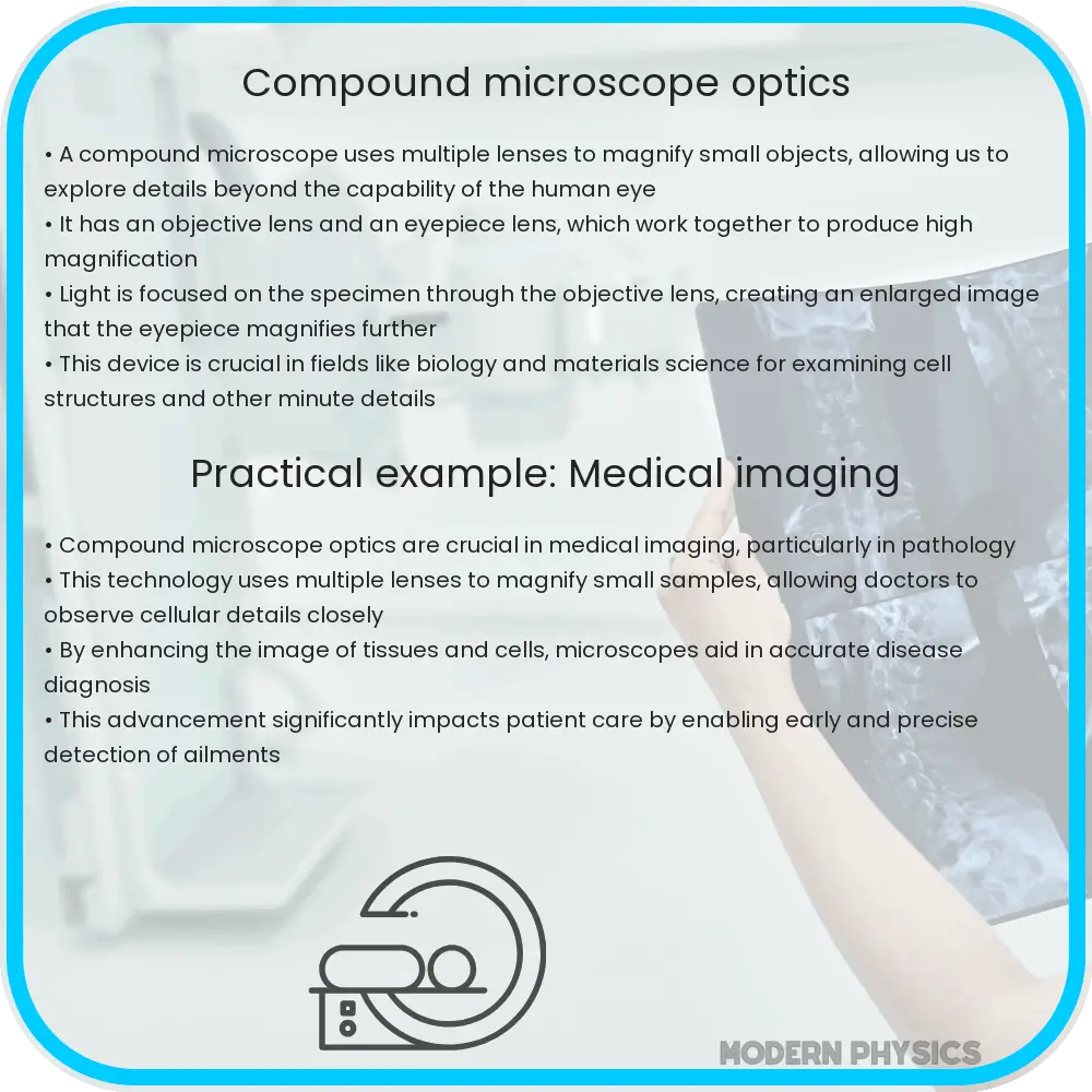
At the heart of a compound microscope are two types of lenses: the objective and the eyepiece. The objective lens, situated close to the specimen, collects light and creates an enlarged image of the object. This image is then magnified further by the eyepiece lens, where it can be viewed by the observer. The combination of these lenses allows for high levels of magnification, typically ranging from 40x to 1000x.
Precision and Clarity
The precision and clarity of an image produced by a compound microscope depend on several factors. The resolution, or the ability to distinguish two points as separate, is critical for clarity. This is influenced by the numerical aperture of the lenses and the wavelength of light used. Higher numerical apertures and shorter wavelengths of light improve the microscope’s resolving power.
Magnification and Its Limits
Magnification is a measure of how much larger a microscope can make an object appear. However, increasing magnification without considering resolution can lead to empty magnification, where the image becomes larger but not clearer. The optimal magnification provides an enlarged image that maintains detail and clarity, which is essential for accurate scientific observation and analysis.
Understanding the optical principles behind the compound microscope’s design, such as light refraction and lens curvature, allows for the optimization of these devices for specific applications. Whether for educational purposes, medical diagnosis, or research, the compound microscope’s ability to provide high precision, magnification, and clarity makes it an indispensable tool in the exploration of the microscopic world.
Understanding Compound Microscope Optics
The compound microscope, a staple in scientific research, offers an intricate view of the microscopic world, magnifying objects beyond the capability of the naked eye. Its ability to provide high precision, magnification, and clarity has made it an indispensable tool in laboratories worldwide. This article delves into the optical principles that enable compound microscopes to reveal the minute details of specimens, from biological cells to chemical crystals.
The Role of Precision in Microscopy
Precision in microscopy refers to the microscope’s ability to produce sharp, distinct images that are true to the specimen’s structure. This precision is achieved through a combination of high-quality lenses, precise mechanical components, and advanced optical techniques. The alignment of the optical components, particularly the objective and eyepiece lenses, is critical for minimizing aberrations and enhancing image clarity.
Magnification Levels and Limitations
Magnification is a measure of how much larger a microscope can make an object appear. Compound microscopes typically offer magnification levels ranging from 40x to 1000x, achieved through the combined powers of the objective and eyepiece lenses. However, magnification without clarity is of little use. Therefore, the microscope’s design ensures that increased magnification does not compromise image quality. The resolution, or the ability to distinguish between two closely spaced points, is ultimately limited by the wavelength of light, as described by the Abbe diffraction limit.
Clarity and Contrast Enhancement Techniques
Clarity in microscopy is not solely dependent on the optical quality of the lenses but also on the use of various techniques to enhance contrast. Techniques such as staining, phase contrast, and differential interference contrast (DIC) microscopy allow for the visualization of transparent specimens by enhancing differences in refractive index or thickness. Fluorescence microscopy takes advantage of fluorescent dyes that emit light when excited, providing detailed images of specific components within a sample.
Conclusion
The compound microscope’s ability to provide high levels of precision, magnification, and clarity is rooted in its sophisticated optical design and the application of various imaging techniques. Through the careful selection and combination of lenses, along with the use of contrast-enhancing methods, researchers can explore the microscopic world in unprecedented detail. As optical technology continues to advance, the potential for discovering new insights into the cellular and molecular realms expands, highlighting the enduring importance of the compound microscope in scientific exploration.
Is this conversation helpful so far?