
Explore the Zernike Phase Contrast Method in optics: enhancing microscopy to visualize transparent specimens with unparalleled clarity and detail.
Zernike Phase Contrast Method: Revolutionizing Optical Microscopy
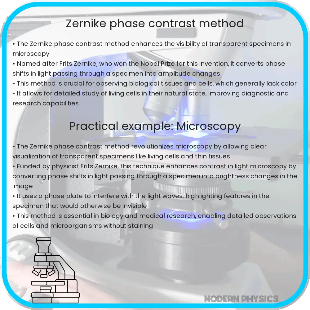
The Zernike Phase Contrast Method, named after the Dutch physicist Frits Zernike who invented it in the early 20th century, stands as a pivotal advancement in optical microscopy. This innovative technique significantly enhances the contrast of transparent specimens, enabling the detailed observation of biological tissues, cells, and subcellular components that are otherwise difficult to see with traditional microscopy methods. Its principle lies in the manipulation of phase differences caused by light passing through different parts of a specimen.
Understanding the Basics of Phase Contrast
At its core, the Zernike Phase Contrast Method relies on the principle that light waves undergo a phase shift when passing through materials of varying thickness, density, or refractive index. These phase shifts, however, are invisible to the human eye because they do not alter the light’s intensity. Zernike’s method ingeniously converts these phase shifts into amplitude differences, thereby making them visible as variations in brightness and contrast in the resulting image.
How It Works: The Role of the Phase Plate
The key component of this method is the phase plate, which is strategically placed in the microscope’s back focal plane. The phase plate is designed to introduce a phase shift between the direct (unscattered) light and the diffracted (scattered) light passing through the specimen. By doing so, it amplifies the contrast between different parts of the specimen, making transparent structures visible and distinguishable under the microscope.
Applications in Biological and Material Sciences
The Zernike Phase Contrast Method has found extensive applications in both biological and material sciences. In biology, it is indispensable for examining living cells and tissues, observing cellular processes in real time without the need for staining or other preparations that may alter or damage the specimens. In the field of materials science, it aids in the analysis of polymers, thin films, and fibers, providing crucial insights into their structure and properties.
Enhancing Detection and Analysis
By converting phase differences into amplitude variations, the Zernike Phase Contrast Method significantly enhances the detection and analysis of specimens in optical microscopy. It allows scientists to observe fine details and subtle changes within samples, facilitating advancements in research and development across various scientific disciplines.
Advantages and Limitations
The Zernike Phase Contrast Method offers several advantages over traditional microscopy techniques, including the ability to visualize transparent specimens without the need for staining or other preparatory methods that could potentially harm living cells or alter their behavior. This non-invasive approach is particularly beneficial for studying biological processes in real-time, providing a clearer understanding of cellular dynamics and functions.
However, there are also limitations to this method. One of the primary challenges is the halo effect, a bright outline that can appear around the edges of specimens, which may obscure fine details. Additionally, phase contrast microscopy is more complex and expensive than basic light microscopy, requiring specialized equipment and phase plates tailored for specific magnifications and objectives.
Technological Advancements and Future Directions
Recent advancements in optical microscopy and digital imaging are expanding the capabilities of the Zernike Phase Contrast Method. Modern phase contrast microscopes often incorporate adjustable phase plates and advanced image processing software, enabling researchers to minimize artifacts like the halo effect and improve image quality. Furthermore, the integration of phase contrast with other imaging techniques, such as fluorescence microscopy, opens new avenues for multi-dimensional imaging of specimens, enhancing both structural and functional analysis.
Looking ahead, the ongoing development of more sophisticated phase plates and computational methods promises to further refine the Zernike Phase Contrast Method. These improvements aim to increase resolution, reduce artifacts, and enable more precise quantitative analysis of phase images. As these technologies evolve, they will undoubtedly unlock new potentials in microscopy, enhancing our ability to explore the microscopic world with unprecedented clarity and detail.
Conclusion
The Zernike Phase Contrast Method has revolutionized the field of optical microscopy, offering a powerful tool for enhancing, detecting, and analyzing transparent specimens with remarkable clarity. Despite its limitations, the method’s non-invasive nature and the ability to observe living cells and delicate materials in real-time have made it indispensable across various scientific disciplines. With ongoing technological advancements, the Zernike Phase Contrast Method continues to evolve, promising to expand our understanding of the microscopic world and fueling future discoveries in science and medicine.