
Learn how PET scans use radiotracer technology to monitor and diagnose cancer, neurological disorders, and heart diseases effectively.
Understanding PET Scans: A Therapeutic Monitoring Guide
Positron Emission Tomography (PET) scans represent a vital tool in the medical field, especially in the diagnosis and monitoring of diseases such as cancer, neurological disorders, and heart diseases. This technology not only aids in detecting the presence of a disease but also provides invaluable information about its stage and response to treatment. In this article, we will delve into how PET scans work and their pivotal role in therapeutic monitoring.
How PET Scans Work
PET scans utilize radioactive substances known as radiotracers to visualize and measure changes at the cellular level within an organism. Typically, a radiotracer such as fluorodeoxyglucose (FDG), which is similar to glucose, is injected into the patient’s body. Since cancer cells consume glucose at a higher rate than normal cells, they absorb more of the FDG. The radiotracers emit positrons, which upon encountering electrons, result in the production of gamma rays. These gamma rays are then detected by the PET scanner to produce detailed images showing the metabolic activity in tissues.
The primary equation governing this positron-electron annihilation process can be expressed as:
E = mc2
Where E represents the energy released, m indicates the mass, and c stands for the speed of light in a vacuum. This fundamental principle by Albert Einstein underpins the high-energy physics involved in PET scans.
Applications in Therapeutic Monitoring
PET scans are particularly valuable in monitoring the effectiveness of cancer treatment. By comparing scans taken over time, doctors can determine if a tumor is responding to specific therapies, such as chemotherapy or radiation therapy. This is crucial for adjusting treatment plans to enhance their effectiveness and potentially reduce side effects.
- Detection of Metabolic Changes: PET scans can detect changes in the cellular activity of tissues, which often occur before physical changes become apparent. This early detection can be a predictor of how well the tumor is responding to treatment.
- Measurement of Treatment Effectiveness: By measuring the intensity of radiotracer uptake before and after treatment, clinicians can assess the efficacy of a therapy, providing a quantitative basis to either continue, modify, or halt a particular treatment regimen.
- Guidance for Biopsy: PET scans help in pinpointing the most metabolically active area of a tumor, which can guide surgeons during biopsy procedures to ensure samples are taken from the most relevant areas.
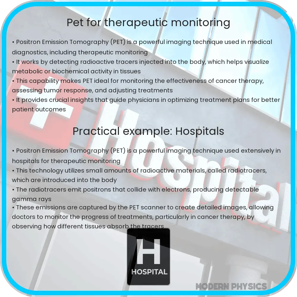
Aside from oncology, PET scans are also employed in the fields of cardiology and neurology. In heart diseases, PET scanning can assess myocardial perfusion and viability, which is essential for diagnosing heart conditions and planning interventions. In neurology, it aids in examining brain functions and diagnosing conditions such as Alzheimer’s disease, Parkinson’s disease, and epilepsy, providing critical information about brain activity and functioning.
In conclusion, PET scans serve as a dynamic and non-invasive method to not only diagnose and monitor various diseases but also tailor treatments according to the unique metabolic behavior of an individual’s condition. As technology advances, the precision and utility of PET scanning continue to improve, opening new avenues in personalized medicine.
Challenges and Future Directions in PET Scan Technology
Despite the numerous benefits of PET scans, there are challenges that limit their widespread use. One major issue is the cost associated with producing and distributing radiotracers, as they often require a nearby cyclotron and a specialized facility for synthesis. Additionally, the short half-life of commonly used radiotracers necessitates quick scanning protocols and effective coordination within clinical settings.
Another concern is the exposure to radiation, albeit small, which still necessitates careful consideration, especially in repeated scans for therapy monitoring. Technological advancements are ongoing to develop new radiotracers with longer half-lives and less radiation risk, enhancing PET scan safety and accessibility.
Technological Innovations Enhancing PET Scans
Recent technological innovations have led to improvements in PET scan imaging and functionality. The integration of PET with other imaging modalities, such as Magnetic Resonance Imaging (MRI) and Computed Tomography (CT), has resulted in systems like PET/MRI and PET/CT. These combinations provide more comprehensive diagnostic information by correlating metabolic activity with anatomical structures, offering superior image resolution and diagnostic accuracy.
Furthermore, advancements in digital technologies and artificial intelligence are revolutionizing the way PET scan data is analyzed. Machine learning algorithms can now process vast amounts of imaging data more rapidly and with greater precision, potentially improving the identification and characterization of diseases.
Conclusion
PET scans epitomize a remarkable fusion of physics, chemistry, and medicine, presenting a critical imaging technique that transcends mere diagnosis to actively guide and monitor treatment across various medical disciplines. While challenges such as cost and radiation exposure persist, ongoing research and technological advancements promise to enhance the capabilities and reduce the limitations of PET scans.
As we look forward, the integration of PET technology with emerging digital tools and more sophisticated imaging combinations continues to refine personalized treatment plans. This not only maximizes therapeutic efficacy but also minimizes unnecessary treatments and their associated side-effects. PET scans are indeed a cornerstone in the evolving landscape of precision medicine, with their full potential yet to be completely realized in clinical practice.