
Myocardial Perfusion Imaging (MPI) is a diagnostic technique used to assess blood flow to the heart and detect coronary artery disease.
What is Myocardial Perfusion Imaging?
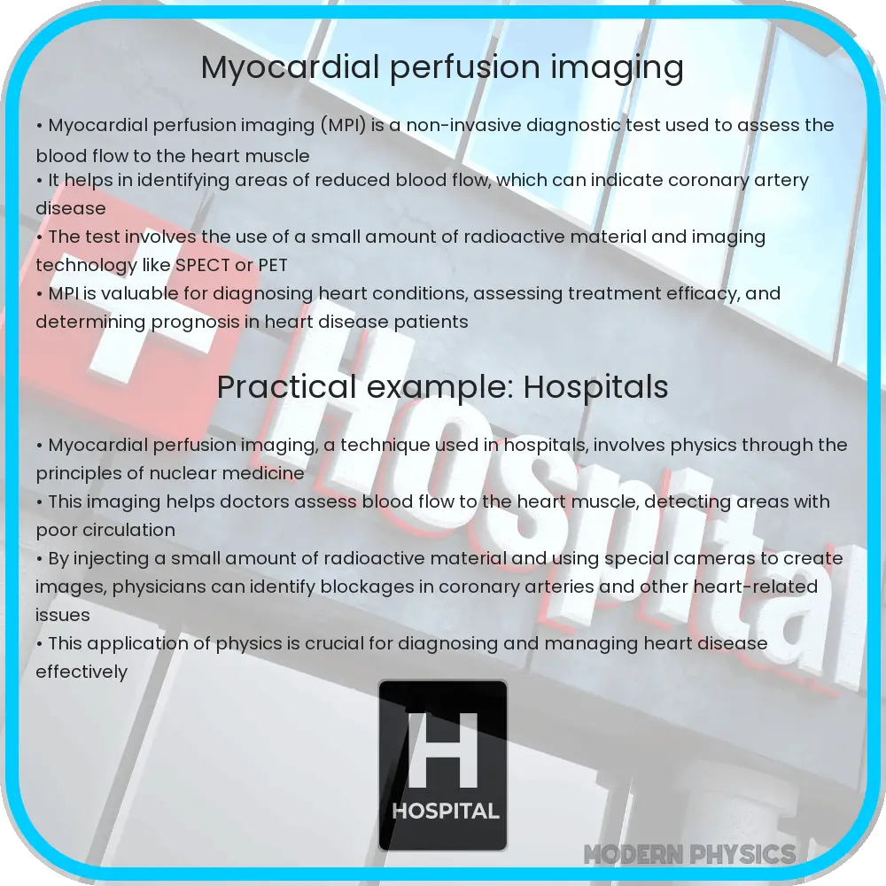
Myocardial Perfusion Imaging (MPI) is a non-invasive diagnostic technique used in cardiology to assess the blood flow to the heart muscle (myocardium). This imaging method helps detect areas of reduced blood flow which can indicate coronary artery disease or other conditions. MPI is often conducted using a nuclear medicine approach, wherein a small amount of radioactive material is injected into the bloodstream.
How Does MPI Work?
During MPI, a radioactive tracer such as Technetium-99m or Thallium-201 is injected into the patient’s bloodstream. As this tracer circulates, it emits gamma rays that are captured by a special camera, creating images of the heart. The procedure is usually performed under both rest and stress conditions to compare the blood flow to the heart muscles while at rest and during exertion (or under induced stress pharmacologically). This comparison helps to identify any discrepancies in blood flow that typically suggest coronary blockages or ischemic heart disease.
Key Uses of Myocardial Perfusion Imaging
- Diagnosis of Coronary Artery Disease: MPI is highly effective in diagnosing coronary artery disease (CAD). It can indicate blockages in the coronary arteries and the extent of any heart muscle damage or scarring.
- Assessment of Heart Attack: After a heart attack, MPI can be used to determine areas of the heart that have been damaged and to assess the effectiveness of treatments.
- Evaluation of Treatment: Doctors often use MPI to evaluate the success of procedures like angioplasty or bypass surgery, ensuring that restored blood flow is adequate post-treatment.
- Risk Stratification: MPI helps cardiologists identify which patients are at higher risk of heart attacks or other cardiovascular events, aiding in deciding the most appropriate therapeutic interventions.
Benefits of Myocardial Perfusion Imaging
One of the major advantages of MPI is its ability to provide comprehensive information about the functional status of the heart in a non-invasive manner. Here are some key benefits:
- Non-Invasive Procedure: Unlike some other procedures that require surgical intervention, MPI is non-invasive, making it less risky for patients.
- Detailed Visualization: MPI offers a clear visualization of blood flow in and around the heart, which can be crucial for accurate diagnosis and treatment planning.
- Effective Monitoring: It allows for the ongoing monitoring of patients with known heart conditions to ensure that their treatment remains effective over time.
- Accessibility: As a widely used procedure, MPI is accessible in many hospitals and diagnostic centers, making it a convenient option for patients.
Potential Challenges and Considerations
While MPI is a valuable tool in cardiology, there are several considerations and challenges that professionals and patients should be aware of:
- Exposure to Radiation: As MPI involves the use of radioactive tracers, there is a small risk associated with exposure to radiation. However, the amount used is typically low and considered safe for most patients.
- Accuracy under Certain Conditions: In some cases, such as in patients with severe obesity or lung disease, the accuracy of MPI can be affected.
- Allergic Reactions: Rarely, patients might have an allergic reaction to the tracer used. It is important to discuss allergies with a healthcare provider prior to undergoing MPI.
- Cost and Availability: The cost of MPI can be high, and its availability may vary depending on geographical location and healthcare facilities.
Conclusion
Myocardial Perfusion Imaging is a crucial tool in modern cardiology, helping doctors diagnose and manage conditions such as coronary artery disease and heart attacks. By providing detailed images of blood flow to the heart under various conditions, MPI assists in making informed decisions about patient care. Despite its several benefits, including being a non-invasive method and offering detailed visualization, it is important for patients to be aware of the potential risks such as radiation exposure and to discuss any concerns with their healthcare provider before undergoing the procedure. Overall, MPI remains a pivotal part of cardiac diagnostics, significantly contributing to improved patient outcomes in cardiovascular care.