
Understanding brain neurotransmitter imaging, a key field in neuroscience that visualizes neurotransmitter activity in the brain to diagnose and treat neurological disorders.
Overview of Brain Neurotransmitter Imaging
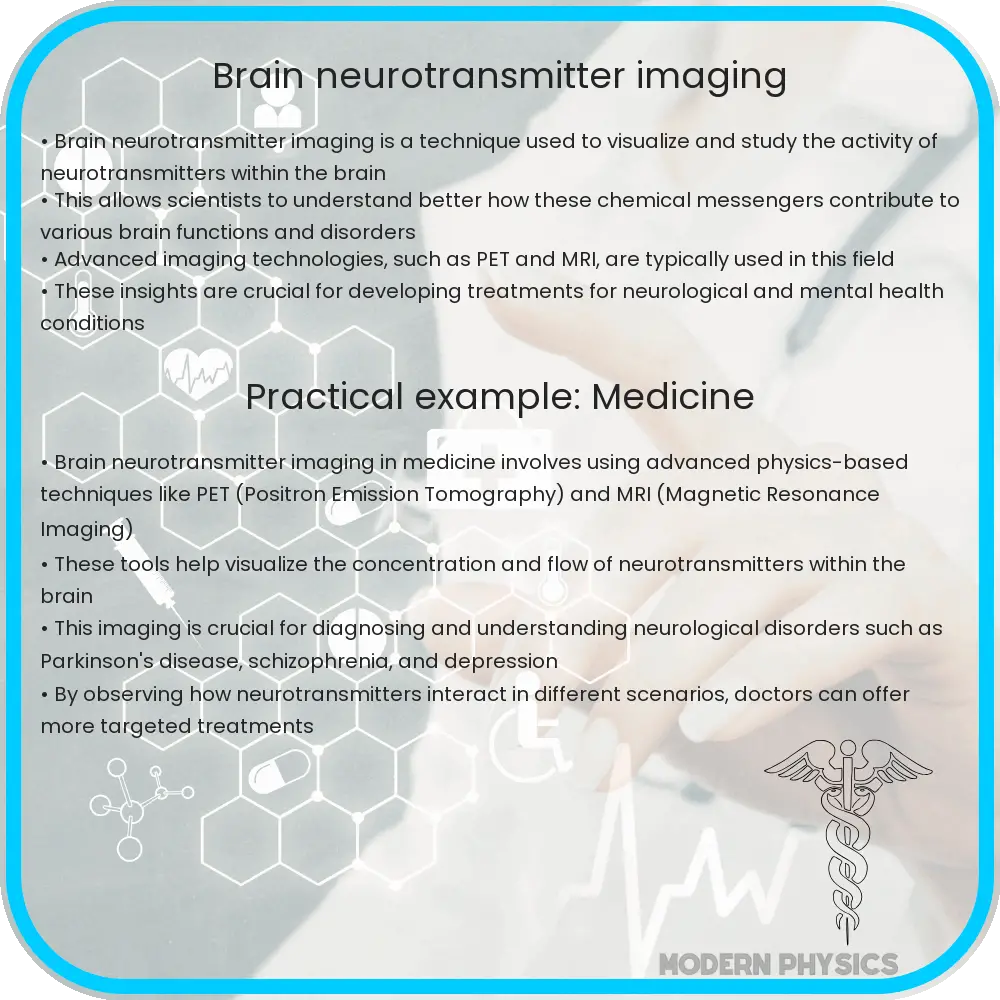
Brain neurotransmitter imaging is a fascinating and rapidly advancing field within neuroscience and medical imaging, aimed at visualizing and understanding the complex interactions of neurotransmitters in the brain. Neurotransmitters are chemical messengers that facilitate communication between neurons, influencing everything from mood to motor control. This imaging helps in diagnosing and understanding neurological disorders, such as Parkinson’s disease, schizophrenia, and depression.
Key Techniques in Brain Neurotransmitter Imaging
Several sophisticated techniques have been developed to image neurotransmitters and their activity in the brain, each with its unique advantages and limitations. Here are some of the main methods currently in use:
- Positron Emission Tomography (PET): PET is a highly sensitive technique that uses radioactive tracers designed to bind to specific neurotransmitter receptors or enzymes involved in neurotransmitter metabolism. By detecting the gamma rays emitted by these tracers, PET can provide dynamic images of the biochemical processes taking place in the brain.
- Single Photon Emission Computed Tomography (SPECT): Similar to PET, SPECT also uses radioactive tracers and gamma rays. However, SPECT tracers emit single photons as opposed to the positron emitters used in PET. The images from SPECT are generally less detailed than those obtained from PET, but the technique remains useful due to its lower cost and wider availability.
- Magnetic Resonance Spectroscopy (MRS): MRS is a non-invasive imaging technique that can be used to measure the concentrations of various neurotransmitters in different parts of the brain without the use of radioactive tracers. It works by detecting the unique chemical signatures of different substances through the use of powerful magnetic fields.
Each of these techniques allows scientists and doctors to visualize and measure different aspects of neurotransmitter activity in the brain, providing crucial insights not only for medical diagnosis but also for the treatment of various neurological conditions.
Detailed Insight into PET and SPECT Technologies
Both PET and SPECT are nuclear medicine imaging techniques that offer a view into the ongoing biochemical processes by tracking specially designed radioactive substances known as radiotracers. In PET imaging, a biologically active molecule, such as glucose, is labeled with a positron-emitting radioisotope. When these positrons meet electrons in the brain, they annihilate each other and produce gamma rays which are then detected by the scanner to produce a detailed image of the brain’s metabolic activity.
SPECT imaging, while conceptually similar to PET, uses different isotopes that emit single photons detectable by the gamma camera. Although SPECT provides lower resolution images compared to PET, it is more readily available and can be used effectively to monitor chronic conditions, assess brain perfusion, and more.
Challenges and Future Directions in Neurotransmitter Imaging
Despite the advances in imaging techniques, there are several challenges faced by researchers and clinicians in the field of brain neurotransmitter imaging. One major challenge is the limited availability of specific radioligands that can bind selectively to certain neurotransmitters without affecting others. Additionally, the cost and accessibility of high-resolution imaging technologies remain significant obstacles, particularly in low-resource settings.
Looking forward, the development of new radioligands and improvements in imaging technology promise to enhance the resolution and specificity of neurotransmitter imaging. Moreover, integrating these imaging techniques with other forms of neurological studies, such as genetic profiling and behavioral analysis, could lead to a more comprehensive understanding of brain function and disorders.
Conclusion
Brain neurotransmitter imaging stands as a critical pillar in the understanding of how our brains function and the pathophysiology of various neurological diseases. Techniques like PET, SPECT, and MRS have opened new avenues for diagnosing and treating disorders by allowing a unique glimpse into the chemical processes of the brain. The continual improvement and integration of these technologies are essential for the future of neurological research and patient care.
As researchers overcome current challenges and continue to innovate, we can anticipate more precise diagnostics and personalized treatments becoming a reality, fundamentally changing the landscape of neurology and psychiatry. This would not only help in managing diseases but also in providing a deeper insight into the complex network of neurotransmitters that play a crucial role in defining human behavior and cognitive functions.